/Acute%20Otitis%20Media%20-%20Perforation%2C%202.webp)
/Acute%20Otitis%20Media%20-%20Perforation%2C%201.webp)


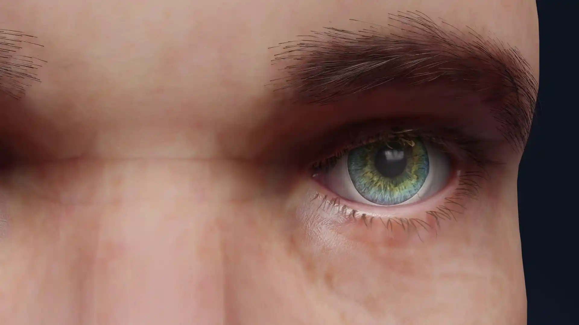
Normal eye image - 30113
Ophthalmology

Select license
More information
Details
Description
This detailed anatomical image showcases a normal human eye surrounded by the orbital structures. The illustration highlights the eyeball centrally encased in layers of extraocular muscles, which control eye movement. The superior and inferior orbital fat pads are also visible, providing cushioning and support within the bony orbit. The clarity of the iris and pupil detail emphasizes the optical axis, while the surrounding soft tissues demonstrate how delicately the eye is positioned and protected within the skull. This educational visual serves as an excellent reference for studying orbital anatomy, including muscle arrangement, fat distribution, and the functional positioning of the eye.
Related items


Item successfully added to the cart