Pterygium stage I image

After purchasing you’ll get a link to a high-resolution file. Your image or video won’t contain watermarks.

Description

This detailed anatomical image illustrates Stage I of pterygium. The lesion is seen as a slightly raised, translucent area extending from the nasal conjunctiva toward the limbus. At this early stage, the pterygium is confined to the conjunctiva and has not yet reached the central visual axis, posing minimal interference with vision. Surrounding anatomical features, including orbital fat and extraocular muscles, provide essential context for understanding the eye’s structural relationships. This visualization is ideal for educational use, highlighting early pathological changes and guiding diagnostic awareness.

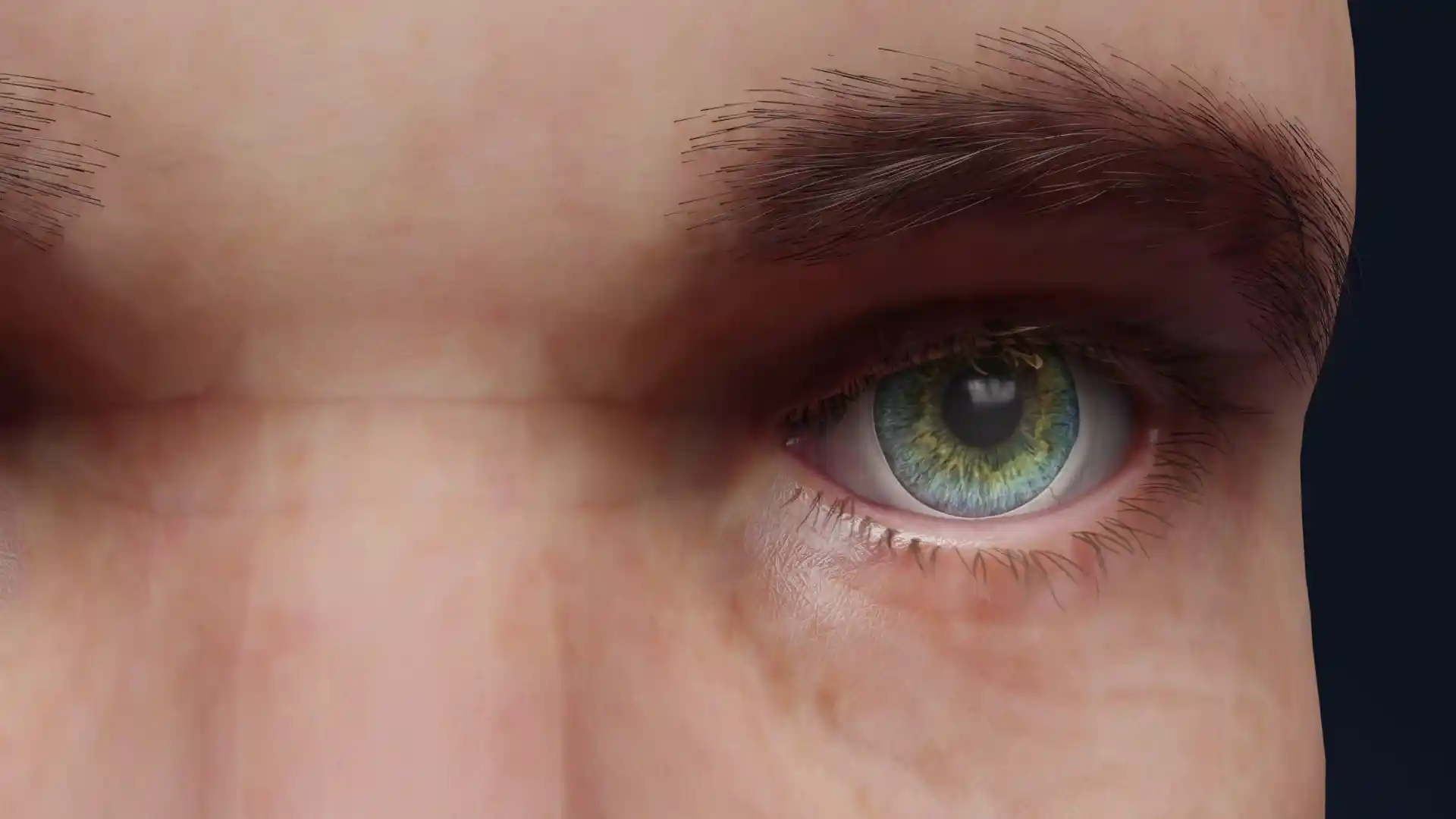
Pterygium stage I image - 30114

Ophthalmology

    License

    Commercial license grants the right to use an image for commercial purposes, such as advertising and marketing.

    Editorial license allows use for news, commentary, or non-commercial publications.

    Personal license grants permission for personal use, creating personal items like wall art.

    Print license authorizes the use of an image in physical print media, such as books, magazines, brochures, and posters.

    -Print

    Quantity

    Specify how many items you need. Bulk orders decrease the cost per unit.

    -More than 50

    Additional information

    Please be informed that when you click the Send message button VOKA Shop will process your personal data in accordance with our Privacy Policy.

    Pterygium stage I image

    Select license

    More information

    Details

    Background N/A
    Resolution 1920 x 1080 px
    Orientation Horizontal
    Format PNG
    File size 2.1 Mb
    Upload date June 10, 2025

    Item successfully added to the cart

    Link successfully copied to clipboard

    Thank you!

    Your message is sent!
    Our experts will contact you shortly. If you have any additional questions, please contact us at shop@voka.io