/Acute%20Otitis%20Media%20-%20Perforation%2C%202.webp)
/Acute%20Otitis%20Media%20-%20Perforation%2C%201.webp)


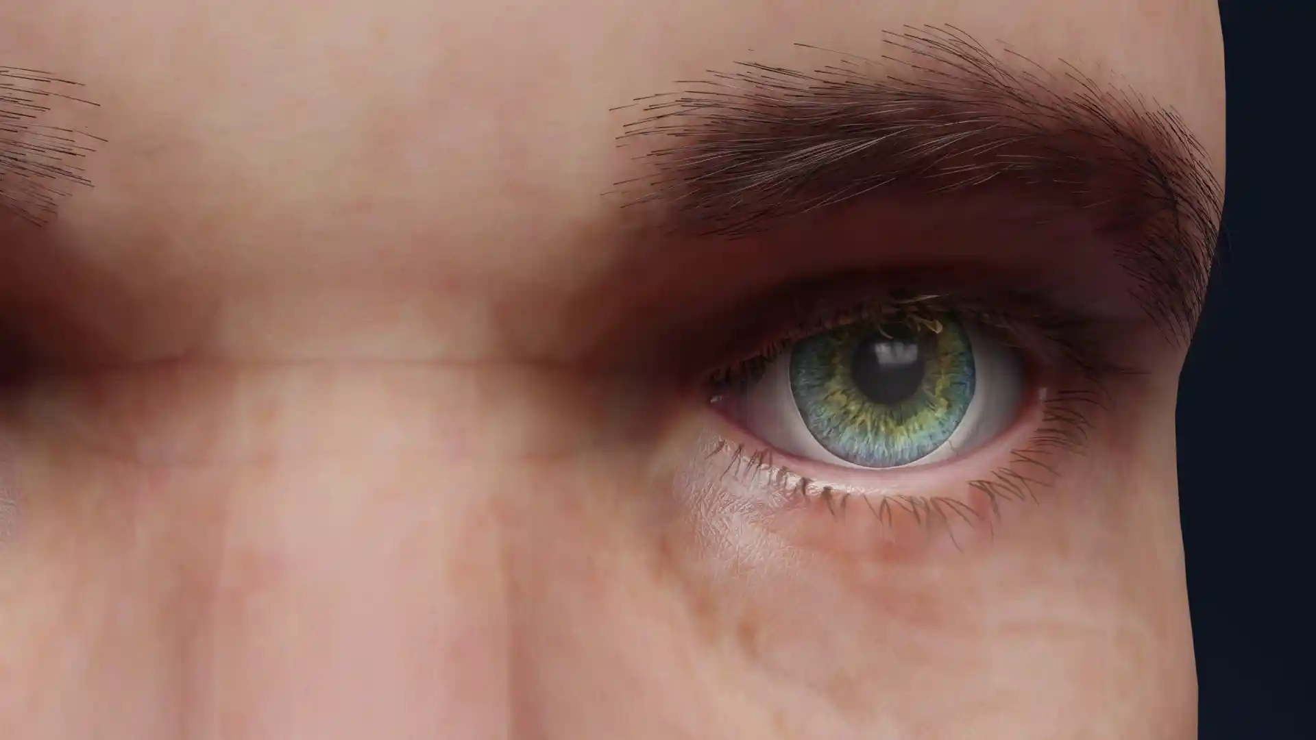
Pterygium stage III image - 30116
Ophthalmology

Select license
More information
Details
Description
This detailed image represents Stage III pterygium, characterized by a fibrous-vascular growth that has advanced well into the central cornea, approaching or overlapping the visual axis. The lesion appears thickened and opaque, with pronounced vascularization and significant corneal invasion, which may cause notable visual impairment. The surrounding structures, including the fatty orbital tissue and ocular muscles, provide anatomical context for surgical planning and education. This stage often requires surgical intervention due to the high risk of vision obstruction. The visualization is a valuable tool for clinicians and educators in illustrating the severe progression and anatomical implications of advanced pterygium.
Related items


Item successfully added to the cart